How does bioprinting work? The field uses printers to place cells and materials layer by layer. Engineers design digital models. Technicians load living cells into bioinks. Printers deposit the bioinks to build tissue structures. Scientists then mature the printed parts in controlled environments. Clinicians plan how to use the tissue in tests or therapies. This guide explains core principles, common methods, bioink makeup, and post-printing steps.
Key Takeaways
- Bioprinting works by depositing bioinks containing living cells layer by layer according to digital models, enabling the creation of organized tissue structures.
- Inkjet and extrusion bioprinting are common methods that differ in bioink viscosity compatibility and cell deposition techniques, balancing resolution and cell viability.
- Advanced techniques like vat polymerization and laser-based methods achieve higher precision but require photosensitive materials and careful cell protection.
- Bioinks are carefully prepared mixtures of cells, carriers like gelatin or alginate, and additives that ensure printability and biological support for cell growth.
- Post-printing maturation involves nurturing printed tissues in controlled environments, with vascularization strategies like hollow channels or endothelial cells to support tissue thickness and viability.
- Bioreactors play a crucial role in tissue development by providing nutrients and mechanical stimuli, while integration with host systems requires careful matching of immune and mechanical properties.
The Basic Principles Of Bioprinting
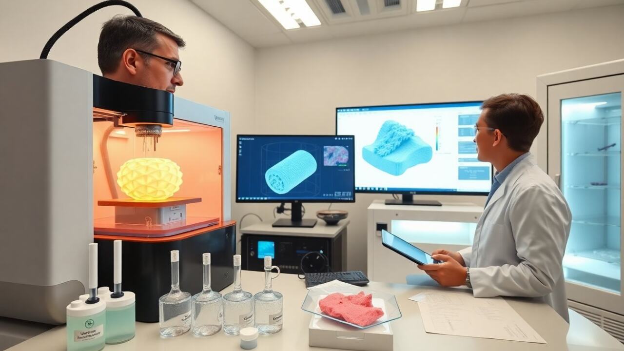
Bioprinting works by converting a digital model into physical tissue. Software slices the model into layers. The printer reads each layer and deposits bioink where needed. Cells, supporting polymers, and signals occupy the bioink. The printer controls placement, speed, and shape. Temperature and humidity affect cell survival during printing. The goal is to place cells so they form organized tissue after maturation. Quality control checks cell viability, print fidelity, and material consistency. Researchers repeat small tests before larger prints.
Common Bioprinting Technologies
Labs use several core printing technologies. Each method changes how printers deposit cells and materials. Choice depends on resolution, cell type, and material viscosity. The next subsections explain two main groups and their trade-offs.
Inkjet And Extrusion Bioprinting: How They Deposit Cells
Inkjet bioprinters eject droplets of bioink onto a substrate. The printer controls droplet size and placement. Inkjet suits low-viscosity bioinks and high speed. Extrusion bioprinters push continuous filaments of bioink through a nozzle. The printer controls pressure and nozzle diameter. Extrusion suits higher-viscosity gels and dense cell suspensions. Both methods keep cells alive when operators set gentle parameters. Labs check shear forces and nozzle clogging during setup. Teams balance resolution, throughput, and cell health.
Vat Polymerization And Laser-Based Methods: Precision And Resolution
Vat polymerization exposes a liquid resin with light to solidify precise shapes. The printer moves light to cure thin layers. Laser-based methods use focused beams to place cells or to trigger material changes. These methods provide higher resolution than droplet or extrusion systems. The methods require photosensitive materials or special cell-handling steps. Teams protect cells from excessive light or heat during processing. Researchers select these methods for small, detailed structures like microchannels.
Bioinks: Composition, Preparation, And Functional Additives
Bioinks combine cells with a carrier material and additives. Common carriers include gelatin, alginate, collagen, and synthetic hydrogels. Scientists suspend target cells in the carrier at defined concentrations. Teams add growth factors, adhesion peptides, or crosslinkers to guide cell behavior. Labs adjust viscosity so printers can deposit the bioink cleanly. Preparation includes sterilization, mixing at controlled temperature, and viability checks. Researchers test each batch for pH, osmolarity, and cell survival. Good bioink design balances printability with biological support for cells.
Post-Printing Maturation And Strategies For Vascularization
After printing, tissue requires time to gain function. Teams place prints in culture with nutrients and oxygen. Cells remodel the carrier and form extracellular matrix. Without blood vessels, diffusion limits tissue thickness. Researchers use several vascular strategies. One strategy prints hollow channels to carry fluid. Another strategy mixes endothelial cells that form vessel networks. Teams also use growth factors to induce vessel growth. Researchers test perfusion to confirm channel function. The aim is to produce thicker tissue that survives after implantation or testing.
Bioreactors, Cell Culture, And Integrating Printed Tissue With Host Systems
Bioreactors supply controlled flow, nutrients, and mechanical cues to prints. Technicians set flow rates, temperature, and gas levels in the reactor. The conditions encourage cells to align, mature, and form functional tissue. Teams monitor markers of differentiation and mechanical strength. For integration, surgeons or researchers assess immune compatibility and graft stability. They use scaffolds or adhesives to connect printed tissue to host structures. Preclinical tests measure vascular integration and function over time. Successful integration requires matching mechanical properties and immune considerations.